Dressage is supposed to be a series of gymnastic exercises that makes the horse healthier, sounder… why then are there so many problems with lameness? Dr Robin Bell looks at the problem…
What is required of a dressage horse?
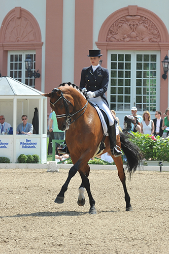
The FEI rules state that the object of dressage is the harmonious development of the horse into an athlete. The result of this development is a horse that is calm, supple, loose and flexible, but also confident, attentive and keen. Dressage is the ultimate competition for many horses and their riders and its popularity continues to escalate. It is considered by some observers to be the most demanding of all athletic equestrian sports. The horse is required to compete in all the paces as well as perform exacting movements.
The locomotor system must be tuned to perfection and any form of lameness is a serious problem for these horses. Elite horses must have a combination of suppleness, balance and power – coupled with expressive and elegant gaits. Training dressage horses, whether at the lowest level or FEI Grand Prix relies on the use of gymnastic exercise to condition muscles and help minimise the risk of injury to ligaments, tendons.
What stresses are involved in training?
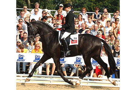
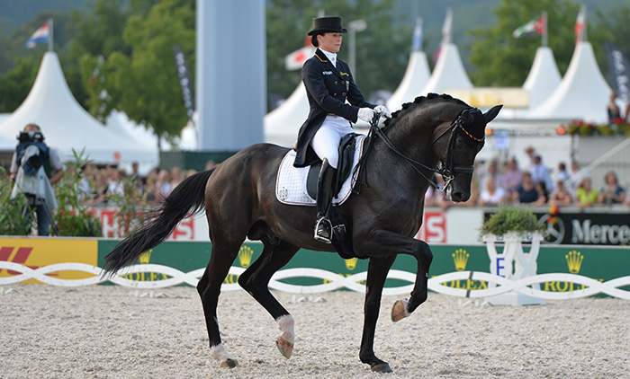
The movements required in dressage tests differ according to the horse’s age and level of training. With lower level tests, the focus is on balance and freedom of movement, medium and advanced levels introduce lateral movements and more collection. The higher classes from Prix St Georges up require maximum collection, impulsion and suspension to enable movements such as canter pirouettes, piaffe and passage. These advanced movements place particular stress on the horse’s skeleton, with lateral movements in particular placing strain on the back and pelvis, and twisting movements on the joints in the limbs. Increased engagement of the hindquarters through collection is needed to perform movements such as passage and piaffe.
Dressage horses are trained on a variety of surfaces, but predominately on sand or artificial footing. Lower level competitions in Australia are still often on grassed arenas, but higher level competitions are almost exclusively held on sand or artificial surfaces. A recent survey of over 11,000 dressage horses in the United Kingdom revealed insights both into the incidence of lameness problems in horses and the possible relationship to arena surfaces. In terms of arena surface, this study illustrated that horses worked on arenas with a sand component had the highest incidence of lameness issues. Conversely there was found to be a slight protective effect when horses were worked for a prolonged period on sand surfaces, most likely due to an adaptation of the bones, joints and soft tissues to the loads experienced when working on this surface. Additional risk factors for lameness in the above study include a change in arena surface when wet, to becoming deep or boggy, and poor arena maintenance.
What types of lameness can we expect to see in dressage horses?
In this part of the article I will outline how to diagnose lameness in dressage horses and to give my perspective on some of the conditions that I see most often in my practice, which deals mainly with sports medicine and performance cases.
Lameness examinations for dressage horses are essentially the same as in other horses, requiring a detailed physical examination, examination in motion, nerve or joint blocks and diagnostic imaging. Often in high performance dressage horses the lameness or clinical complaint may only be visible under saddle, and in more subtle cases may only be perceptible to the rider. Low-grade hind limb lameness in particular may only be visible in certain specific circumstances, such as finding a flying change or canter pirouette more difficult in the direction towards the lame limb, or the half pass more difficult in the direction away from the lame hind limb. It is also useful to keep in mind that while inexperienced riders may make a sound horse appear lame, a competent rider may actually make a subtly lame horse appear sound. This may occur almost subconsciously to the experienced rider and for this reason I often ask these riders to deliberately ride ‘badly’.
Due to the nature of the work we ask dressage horses to perform it is also important to look at the horse as a whole when assessing for lameness, rather than just focusing on the limbs. Subtle changes such as an increase in suppleness through the back or neck after nerve blocks, or a change in engagement of the hindquarters during transitions after analgesia, may indicate that the problem is originating from the region that was blocked. Again due to the subtle clinical signs that are observed, it is also important that the surfaces that the lameness is evaluated on are kept as consistent as possible between nerve blocks.
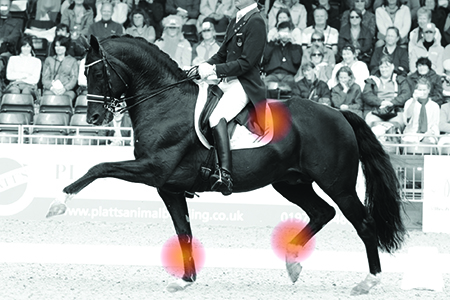
The most common lameness problems encountered in dressage horses are outlined in table 1. In a similar study to that mentioned previously, in UK dressage horses it was shown that a third of almost 2600 horses had a lameness issue during their career. Of these, 50% of Grand Prix horses and approximately 20% of horses at other levels were reported to have a lameness issue in the preceding two years. Over a third of these problems were reported to come from the foot, again reinforcing the adage ‘no foot, no horse’. The next most common regions affected were the suspensory ligament (15%), and the hock region (12%).
Common areas that can cause issues and lameness
This article will focus on three conditions that I encounter most frequently in dressage horses: proximal suspensory desmitis, bone spavin and coffin joint arthritis.
Proximal suspensory desmitis.
Proximal suspensory desmitis (PSD) is an injury to the suspensory ligament at its origin from the top of the cannon bone. It occurs in the hind limbs and the fore limbs.
Hind limb PSD is most common in horses working at medium and advanced levels, and is often seen in both hind limbs at the same time. This can make the detection of the problem difficult for the rider as there may be no overt lameness present, rather just a loss of performance, stiffness, or resistance. Injection of local anesthetic around a specific nerve can help with the diagnosis of this problem. In the hind limbs there may be no pain upon palpation due to the location of the ligament deep to the splint bones. This location also makes diagnosis using ultrasound difficult, and the use of x-rays or bonescan may be necessary to complete the diagnosis. Treatment depends on the severity of the lesions on ultrasound and the chronicity of the condition. Many horses with an acute injury and good conformation respond well to 3 months rest. In horses with chronic injury, the results of even prolonged rest are often disappointing. In such cases I prefer to use injection of anti-inflammatories around the ligament and institute a controlled exercise program. In refractory cases surgical intervention, often combined with therapies such as stem cell or platelet rich plasma injections is the only option.
Forelimb PSD is more common than hind limb in young horses. It is thought that it may result from over-extension of the carpus in extravagantly moving young horses, especially those who volunteer extended trot. The lameness in PSD is more often seen in only one limb, and is often worst with the affected leg on the outside of the circle. The lameness may in some cases only be seen with the horse under saddle. In very acute cases there may be some mild swelling present around the ligament, but this resolves very quickly. PSD in the forelimbs is diagnosed using nerve blocks and ultrasound. Treatment relies on a period of 3-4 months rest and a controlled exercise program of increasing walking. If there are noticeable core lesions present injection of stem cells or platelet rich plasma is useful. I find that with an adequate rehabilitation program, treatment with shockwave therapy most horses with acute lesions can return to full performance. Horses with chronic lesions are prone to re-occurrence.
Lateral movements place strain on the back and pelvis, and twisting movements on the joints in the limbs
Bone spavin
Bone spavin is the colloquial term for osteoarthritis and pain in the distal intertarsal and tarsometarsal joints of the hock. It has been recognized for hundreds of years. Bone spavin may cause overt lameness or poor performance. Horses with this condition may have an expressive free trot, but a poor canter, and in particular have problems in more collected gaits, where there is increased loading of the hock. Advanced horses may show irregularities of rhythm in piaffe or passage. These clinical signs may often be confused with the horse having a sore back. Diagnosis relies on nerve blocks, and x-rays, but it is important to note that there is often a poor correlation between the response to nerve blocks and the severity of radiological changes. I see a lot of horses that have lameness localized to the hock joint but minimal changes on x-rays, in these horses a bonescan is often used to confirm the diagnosis. Most horses respond well to injection of anti-inflammatories into the joint, combined with corrective shoeing. The use of ‘anti-arthritis’ drugs containing pentosan polysulfate may also be of use. Some horses with pain originating from the bone underneath the joints may benefit from treatment with the bisphosphonate tildren. In cases refractory to the above therapy, fusion of the joints either chemically or surgically may result in a return to performance, though often at a lower level than previously.
Coffin joint arthritis
Pain arising from inflammation or arthritis within the coffin joint is a common problem in all performance horses, not just the dressage horse. The location of this joint within the rigid hoof capsule and the forces it is subjected to are thought to be part of the predisposing factors for pain from this region. Poor foot conformation and medial-lateral imbalance are also important contributors to lameness from this region. Diagnosis requires specific nerve blocks including coffin joint blocks, but interpretation of these blocks is not straight forward, and it is easy to have a false positive response. The presence of arthritis or chip fractures on x-rays or evidence of inflammation and excess joint fluid on MRI scans provide confirmation of the diagnosis.
Treatment is a combination of intra-articular medications, most commonly with low dose corticosteroids and hyaluronic acid, or other products such as IRAP or platelet rich plasma. In young horses I tend to recommend the use of IRAP, because of the likelihood that the horse will need repeated injections over the course of its career. Corrective shoeing to improve the foot-pastern axis and correcting any imbalance is essential but should be performed gradually over multiple shoeings. The prognosis for horses without advanced x-ray changes is good, and most horses can return to full work.
With any form of lameness, performance can be seriously impaired. All cases of lameness should be treated appropriately, as well as ensuring that adequate preventative strategies are in place, including preventative medications and excellence in shoeing.
Table 1: Common clinical conditions causing lameness in dressage horses
| Condition | Clinical signs | Diagnostics | Treatment | Prognosis |
| Proximal suspensory desmitis | Loss of performance, change in contact or resistance, problems with pirouettes or flying changes | Lameness exam, nerve blocks, x-rays, ultrasound, MRI or CT | Rest and controlled exercise, ultrasound guided injections of stem cells or growth factors, surgery, shockwave therapy | Fair-good |
| Osteoarthritis of the distal hock joints | Unwillingness to work, toe dragging, shortened stride behind | Lameness exam, nerve blocks, x-rays, bonescan | Corrective shoeing, phenylbutazone and light work, intra-articular injections, joint fusion via chemical or surgical arthrodesis | Good-Excellent |
| Osteoarthritis of the coffin joints | Foot imbalance, shortened stride if both limbs, acute lameness if one limb, | Lameness exam, nerve blocks, x-rays | Corrective shoeing, intra-articular medications, rest and phenylbutazone, joint supplements | Good |
| Osteoarthritis of the fetlock joints | Fetlock joint swelling, loss of range of motion, moderate lameness | Lameness exam, nerve blocks, x-rays | Corrective shoeing if imbalance is present, rest, intra-articular medications, joint supplements | Fair-good, better prognosis if minimal signs of disease on x-rays, and better for forelimbs than hindlimbs |
| Suspensory branch desmitis | Enlarged branch on palpation, painful to palpate, mild-moderate lameness | Lameness exam, nerve blocks, ultrasound | Rest, controlled exercise programme, injection of stem cells or growth factors | Fair-Good, 6-9 months rehabilitation, prone to re-occurence |
| Palmar or plantar annular ligament desmitis | Swelling over the back of the fetlock, moderate lameness that worsens with work, windgalls with a notch over the ligament | Lameness exam, nerve blocks, ultrasound | Rest and rehabilitation, surgical release of the ligament | Good |
| Thoracolumbar pain | Poor performance, lack of suppleness, resentment of prolonged exercise | Lameness exam, nerve blocks, bonescan, x-rays | Rest, physiotherapy and rehabilitation program | Fair-Good |
| Sacroiliac Joint Disease | Unwillingness to perform certain movements, loss of impulsion, stiffness, worse under saddle than on the lunge | Lameness exam, bonescan, ultrasound | Modification of work program, ultrasound guided injection of corticosteroids | Fair |
| Check ligament desmitis | Sudden onset lameness, heat and swelling over ligament in the cannon | Lameness exam, nerve blocks, ultrasound | Controlled exercise with minimal rest | Fair-Good, may be prone to re-occurence |
| Osteochondrosis | Joint swelling mild-moderate lameness | Lameness exam, x-rays | Rest, arthroscopic surgery for removal of bone chips | Fair-Good |
| Tenosynovitis of the Digital Sheath | Chronic windgalls with acute lameness, pain on flexion of fetlock. May be associated with annular ligament desmitis | Physical examination, nerve blocks, ultrasound | Injection of medications into the sheath, tenoscopic surgery (keyhole), controlled exercise | Good if no tendon injuries or adhesions within sheath. |
I’m not sure I understand this sentence: “In terms of arena surface, this study illustrated that horses worked on arenas with a sand component had the highest incidence of lameness issues. Conversely there was found to be a slight protective effect when horses were worked for a prolonged period on sand surfaces, most likely due to an adaptation of the bones, joints and soft tissues to the loads experienced when working on this surface.”
What is the difference between sand component and sand surfaces? Is that what makes a difference?
I do not feel that this article answers the question it asks in the first paragraph!
Perhaps the figures should be compared with those of the Spanish Riding School, is there a difference between modern dressage and what the word dressage really means… to train the horse for maximum ride-ability, well being and longevity!
If people want to do this to their horses then they should call it something different or move away from the over exuberant movement which has become the norm, it is appealing and pretty, gets the marks but is not true dressage.
Excellent question and one that I am asking myself but more importantly I ask myself has my riding been up to standard. Answer, no because my horse has kissing spines and showing mild coffin joint lameness. May be result from him fracturing it as a 3 year old, 14 years ago, but I have done a fair amount of dressage and find that he has these issues. I became too focussed on results in my tests rather than quality riding. You don’t hear the Spanish Riding Schools having these issues. They spend years doing correct training. So I’m following classical in hand work in the hope I can resolve the asymmetrical imbalance that my riding has caused combined with physio and veterinary treatment. Nothng to be proud of – I screwed up my horse so I must put him right and learn to ride correctly even if it means saying stuff competitions!
Cristiana on August 9, 2014 asked the difference between sand component and sand surfaces..
The sand arena is made using plains sand.
The sand component used in the more modern arenas today use a mix of sand, geotextile ans fribers to help keep the sand at proper moisture and to help minimize impact on horses joints.
Why do dressage horses go lame so often? Easy. Compare photos of today’s dressage horses with horses from the 1920s and 1930s. They are not only taller, but also bulkier. The horses of the 1930s were about 1000 lbs. Take a typical dressage horse of today at 1200 to 1400 lbs, and he is carrying 1 1/5 to 1 1/2 more weight than the dressage horses of old, and that’s before you add the rider. Obviously the horse’s own body weight is increasing the stresses on all his joints and tendons.
Secondly, many sport horse riders are great fans of support boots “to protect my investment” they say. Overuse of support boots means the horse’s legs are used to being supported. When the boots come off for the competition the horse’s legs are not getting the usual support. Voila, he’s lame.
I also think it is the type if riding people are doing, as well as the boots and bandages, lack of turn out. Riding something else besides ‘dressage’ might help. Look at all the horses that competed for years and what they did …klimke’s videos show on a track galloping, cavalltti work, jumping.
Horses now, as well as being bigger are also smaller boned.
You should not lay a horse unless you are sure there reaches least one horse which includes
the application to overpower your selection, ideally there
needs to be two or three. Some people really enjoy it although some try to produce
a living from it. Be bound to look out to the conditions before you decide
to decide on a horse racing game to play.
It was interesting some years ago watching Harry Boldt coach a rider while the Australian coach was coaching is the next arena. Mr Boldt have the horse a few excercise then said “walk and let him rest” . After a rest he started the horse again. The Australian coach had the other horse in a lather of sweat and continued to hammer the horse as it became tired. Mean while Mr Boldt has his horse calm relaxed and doing short bursts of brilliance
Top tips for avoiding these injuries would be a useful extension to the article.
I think it is your trainers JOB to teach CORRECT dressage. If you do not have a trainer that can actually correct you, and teach you the long hard road that is training a dressage horse the answer is simple…GET A BEW TRAINER. It is not a question of “modern” or “classical” dressage. It is a question of “correct” and “incorrect”. Much of what scores high is incorrect, but it differs from the actual rules on scoring. Read an article or two by Jeremy Steinberg, he is a breath of fresh air.
I am lucky to have a trainer who has helped me start a very difficult horse. I am happy to say after 4 years under saddle, and schooling PSG we actually RARELY school tricks. We work basics most of the time, because without a foundation you cannot build a house.