Our two veterinary experts, Dr Robin J.W. Bell and Professor Leo Jeffcott (University Veterinary Teaching Hospital Camden and University of Sydney) look at the common problems that showjumpers develop – how to identify them – and how to treat them…
Showjumpers – The TEST
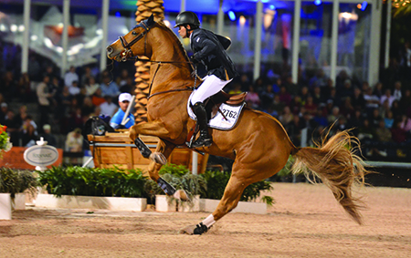
Showjumping is a serious athletic test for both the horse and rider. Horses competing in showjumping are required to jump obstacles from 0.90m to upwards of 1.7m in elite competition at major championships. Not only are the heights a challenge, but the obstacles are presented in combinations at distinct striding intervals to test the horses’ ability to shorten or lengthen their stride on course. The stress is increased in classes against the clock with tight turns placing increased strains on the musculoskeletal system. Most outdoor competitions in Australia take place on grass, but in the USA and Europe there is an increasing tendency for these shows to be on all weather synthetic surfaces.
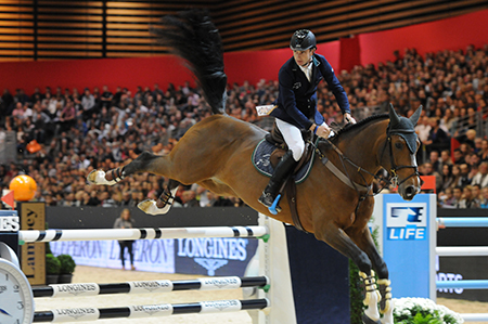
Showjumping is a serious athletic test for both the horse and rider
Problem area 1 – the foot
Because of the tight turns, and variable surfaces, riders often use studs or caulks that are screwed into the shoes to enhance traction. These studs may be placed both on the inside and outside heels of the limbs, but occasionally are placed only in the outside heel of the shoe. If only one stud is used, this can predispose these horses to injury, through foot imbalance and increased torque on the foot/pastern. On hard ground studs have the potential to increase bruising and may predispose these horses to hoof wall cracks.
Problem area 2 – the limbs and joints
On the final approach stride the forelimbs are responsible for breaking the stride and providing vertical force for take-off. This places significant strain on the lower joints in the leg. The hind limbs provide the majority of the force for takeoff, and the head and neck position changes to ensure optimal force generation. The time the hind limb is in contact with the ground on the takeoff stride in the hind limbs is longer than a normal canter stride which allows the horse to generate the forces necessary to successfully clear a fence. On landing the front coffin and fetlock joints are overextended (hyperextended) which places strain on the flexor tendons and the suspensory ligaments in these limbs. It is not therefore surprising that damage to the musculoskeletal system and lameness is so common in showjumping horses.
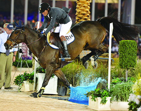
On landing the front coffin and fetlock joints are overextended
Finding the problem:
Performing a lameness examination in showjumpers follows the same protocol as discussed in previous articles, with detailed history, thorough physical examination, nerve blocks and diagnostic imaging as necessary. In addition it may be necessary to examine these horses under saddle over fences, especially in more complex cases. Some subtle injuries can cause a decrease in performance such as drifting to one side when jumping, showing resistance to one rein in particular, difficulty with a flying change in one direction, refusing to jump a certain type of obstacle, rushing at fences, or unwillingness to work on a particular arena surface.
Unlike the studies performed in dressage horses, there are limited published studies on lameness in showjumpers. The largest in showjumpers is a Swedish study which had 231 elite showjumpers from thee European countries. While there were many limitations in this study, the authors showed that horses who had previous orthopaedic injury were much more likely to have time off due to lameness. As with the study in dressage horses, those horses that had variation in their training regimes (i.e. hacking out, different types of work, working on a variety of surfaces) were less likely to have days out of work due to lameness.
Causes of Lameness in Showjumpers
As in previous articles we will concentrate on some of the conditions most often encountered in practice, with a more comprehensive list of the common clinical conditions included in Table 1.
The first of these conditions are lameness associated with structures within the foot. As mentioned in previous articles lameness from the structures within the foot is an extremely common source of lameness in all performance horses. This pain may come from simple causes such as tearing/bruising of the sensitive laminae or hoof wall cracking, or may be related to injuries of the bones/ligaments in the foot like the navicular bone. Jumping horses in particular are prone to injuries in the most distal parts of the deep digital flexor tendon as it passes over the back of the navicular bone and where it inserts onto the coffin/pedal bone. Other common injuries within the foot include damage to the collateral ligaments of the coffin joints, and bruising or trauma to the bones in the foot/pastern (Figure 3). Jumpers are prone to these injuries due to the repeated loading placed onto the coffin joint upon landing after fences. This loading also causes inflammation of the joint itself which may be seen without injury to the soft tissues mentioned above. In Australia a bonescan is commonly used as the main means of attempting to gain a diagnosis in these cases, and while useful in certain cases, it provides only localizing information rather than the specific diagnosis.
MRI: Cost Effective
After the lameness has been localized to the foot region with nerve blocks, the best way to definitively diagnose lameness conditions within the foot is to perform MRI. This is a cost effective means to target therapy to the precise cause of the problem, rather than spending months trying various ‘shotgun’ treatments. Specific treatment depends on the exact diagnosis, simple cases of inflammation within the lining of the coffin joint respond extremely well to medication of that joint with corticosteroids and hyaluronic acid. Soft tissue injuries such as tears in the deep digital flexor tendon or collateral ligaments require a prolonged period of rest, corrective shoeing, and depending on the location of the injury, may benefit from the injection of stem cells or platelet rich plasma directly into the injury. Injuries to the bones of foot/pastern often respond to treatment with the drug Tildren and a shorter period of rest.
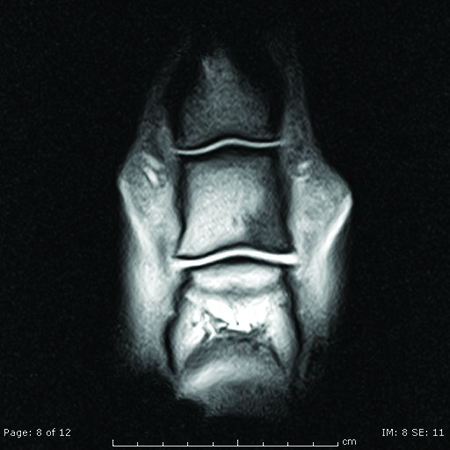
MRI image showing bruising in the short pastern bone (black area)
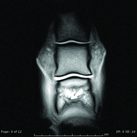
Follow up image at three months showing resolution
Hock Problems
The most common cause of hind limb lameness in jumping horses is arthritis in the bottom joints of the hock (bone spavin). A number of factors can contribute to the development of this condition, ranging from poor conformation, to the extreme strains placed onto the hocks during takeoff. These horses may show a loss of impulsion, and a shortening of stride length. Horses may consistently start to jump towards one side of the fences, and may have more trouble with oxers. Some horses may switch leads in front of a fence or have trouble jumping from one lead or the other. There often is pain upon palpation of the lumbar muscles. A lameness examination will show increased lameness after upper limb flexion tests, and there are commonly changes on x-rays indicative of arthritis. It should be noted that not all horses with clinical signs of hock arthritis have abnormalities on x-rays, and some may need a bonescan and/or nerve blocks to help diagnose the condition. Treatment most commonly involves intra-articular medication with corticosteroids, but corrective shoeing, and the judicious use of a non-steroidal anti-inflammatory such as phenylbutazone can be very helpful. In cases that are not responsive to these treatments, fusion of the joints either surgically or chemically is often very effective. Overall, the prognosis with appropriate therapy is excellent, with the majority of horses able to compete at their previous level of performance.
Because of the athletic demands placed upon them lameness is an extremely common problem in jumping horses, but it does not necessarily mean that the horse’s career is over. Provided an accurate diagnosis is obtained, treatment for most conditions will result in a return to similar levels of performance. As with any performance horse, the use of products that provide some degree of joint protection such as pentosan polysulfate, or hyaluronic acid are indicated on at least a monthly basis.
Extreme strains are placed onto the hocks during takeoff
Table 1: Clinical Conditions causing lameness in the stifle in Showjumping Horses.
| Clinical Condition | Clinical Signs | Work-up | Treatment | Prognosis |
| Arthritis of the bottom hock joints(bone spavin) | Unwillingness to work, dragging toes, shortened stride behind | Lameness examination, nerve blocks, x-rays, bonescan | Corrective shoeing, phenylbutzone and light work, intra-articular injections, joint fusion via chemical or surgical arthrodesis | Good-excellent |
| Suspensory ligament desmitis | Initially subtle, loss of power, or lameness that is warmed out of, lameness worse on soft going or with limb on the outside of the circle | Lameness examination, nerve blocks, ultrasound, x-rays. | Rest and corrective shoeing, graded exercise program, shockwave treatment., injection into ligament of stem cells, PRP, bone marrow, surgical treatment for hindlimb | Fair-good |
| Stifle joint Arthritis | Shortening of stride behind, unwillingness to canter on a certain rein, lameness | Lameness examination nerve blocks, ultrasound, x-rays, diagnostic arthroscopy, bonescan | Intra-articular medications, Stem Cell or PRP injections | Fair-Good |
| Cruciate ligament and meniscal injuries | Often over-diagnosed. Generally moderate-severe hind limb lameness | Lameness examination, nerve blocks, ultrasound, diagnostic arthroscopy | Limited, rest and graded exercise, surgical debridement via key-hole surgery, Stem Cell or PRP injections | Poor-Fair |
| Superficial digital flexor tendinitis | Swelling and pain over the tendons, lameness may or may not be present | Physical examination and ultrasound | Rest and graded exercise program. Intra-lesional injection of PRP or stem cells | Fair-good, prone to re-injury |
| Upward fixation of patella | Locking of stifle if severe, mildly affected horses may show an unwillingness to canter, switching of leads and skipping behind | Lameness examination, ultrasound of the patella ligaments | Increased work-up hills to build gluteal muscles, internal blistering of ligaments with iodine in almond oil, surgery with patella ligament splitting | Excellent |
| Inflammation in the digital sheath | Windgalls, lameness in the affected limb | Lameness examination, nerve blocks, ultrasound | Rest and graded exercise if concurrent injury to tendons, injection of corticosteroids and 1-2 week rest if not, surgery if annular ligament thickened. Keyhole surgery (tenoscopy) if lameness re-occurs | Good-excellent if tendons not injured, fair-good if they are involved. |
| Pastern Joint Arthritis (ringbone) | Stiffness at start of work initially, then moderate lameness in more chronic cases | Lameness examination, nerve blocks, x-rays, bonescan | Intra-articular injections, shockwave, surgical fusion of joint in severe cases | Fair |
| Injury to the oblique and/or straight sesamoidean ligaments | Lameness, pain upon palpation | Lameness examination, nerve blocks, ultrasound | Rest and graded exercise, shockwave therapy | Fair-good |
| Cervical Spine arthritis | Stiffness in the neck, unwilling to bend a certain way, forelimb lameness | Lameness examination, x-rays, bonescan | Ultrasound guided facet joint injections | Good |
8 yr old hanoverian TB X honest jumper doing 1.20 mt classes clear,,,,problems going into canter…changes strides….canter does
not FLOAT..rear quarters are skipping more than cantering…..nave applied strong iodine to shoulders area nr girth that are in pain
….bute before competitions helps,,,,,,,once in competition horse does not stop…..but an effort to warm up in warm up arena….