By Dr Robin Bell and Professor Leo Jeffcott
Equine Performance and Imaging Centre, University Veterinary Teaching Hospital Camden
Sore backs are a very common and often overlooked problem in the performance horse. The soreness may be caused by a primary back problem or it may secondary to an underlying lameness issue, faulty or ill-fitting tack or even inadequate schooling. It must be emphasised that the most common presenting complaint in horses with a back problem is poor performance rather than overt signs of pain in the region.
Diagnosis of back problems can be extremely difficult and must start with a systematic clinical examination. Despite a thorough clinical examination, and even with the use of advanced diagnostic aids, definitive diagnosis of a back injury is often only made after eliminating all other clinical problems.
The diagnostic examination should include a history, including duration and time of onset of clinical signs, the animal’s temperament, and response to previous treatments such as the use of Non Steroidal Anti-Inflammatories (NSAID’s) and manipulation. The horse should be visualised at rest, including palpation and manipulation of the back. Examination should then proceed to walking and trotting in hand and on the lunge, and then examination under saddle. Palpation and manipulation of the back should be repeated after exercise. Kinematic evaluation of gait may also be useful, if the facilities are available.
Radiographs of the affected area can be of value, but require high powered X-ray equipment that is not available to all practitioners. A bonescan (nuclear scintigraphy) is also very useful and is particularly sensitive in diagnosing conditions such as vertebral fractures, osteoarthritis, kissing spines, spondylosis, some sacroiliac problems and stress fractures of the pelvis. It can also diagnose lesions in areas that are unable to be viewed with X-rays alone. Ultrasound examination of the back can yield useful information, particularly in relation to the dorsal sacroiliac ligaments, kissing spines and chronic muscle damage.
Ancillary clinical aids include clinical pathology (blood tests) to check for elevation in muscle enzymes both before and after strenuous exercise. The injection of local anaesthetics into specific areas of the back where pathology is suspected such as around ‘kissing spines’ may also help to relieve clinical signs associated with these conditions and aid in diagnosis.
The judicious use of NSAID’s such as phenylbutazone in a short (2-3 days) course can help to differentiate soft tissue and bony problems as chronic skeletal problems will generally show some temporary improvement, with clinical signs then re-appearing.
CAUSES OF BACK PAIN
Table 1 presents the most common differential diagnoses that may be confused with a genuine back injury. This information is deliberately presented before the clinical conditions affecting the back and spine because it is not uncommon for owners to blame poor performance on a condition in the horse’s spine or back, when in actuality it is due to problems with tack, poor schooling or rider equitation. Additionally the typical signs of a ‘cold back’ seen when initially mounting or tightening a girth are not necessarily an indication of a spinal problem. Hindlimb lameness, in particular bilateral hock issues are probably the most common differential diagnosis, but any clinical condition that affects the horse’s gait can lead to secondary back soreness and stiffness. 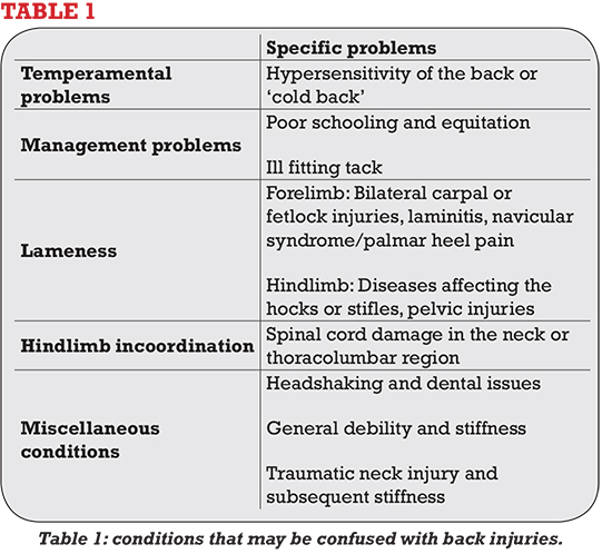
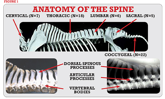
Conditions that may affect the thoracolumbar region
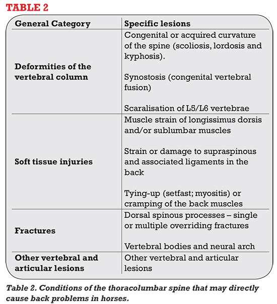
Because of its size and anatomical complexity there are a wide range of problems that can cause back pain and discomfort in the performance horse. These are listed in Table 2.
ALLEGED BACK PROBLEMS
Table 3 lists another category of back problems, which are alleged to cause pain and lameness in the horse. Despite popular opinion there is little anatomical or pathophysiological evidence to support their occurrence. This group of conditions causes much controversy between veterinarians, physical therapists and horse owners. These difficulties are exacerbated by the fact that many horses suffer low grade and chronic lesions. The major clinical sign is always a loss or reduction in performance whatever the underlying pathogenesis; other clinical signs may be more difficult to define. Although the intervertebral discs in horses tend to show degenerative changes with age, they do not seem to cause any clinical signs, unlike the situation in people and dogs. Nerve “pinching” and peripheral nerve lesions are often claimed to be the cause of back problems in the horse, but there is no scientific evidence to support this.
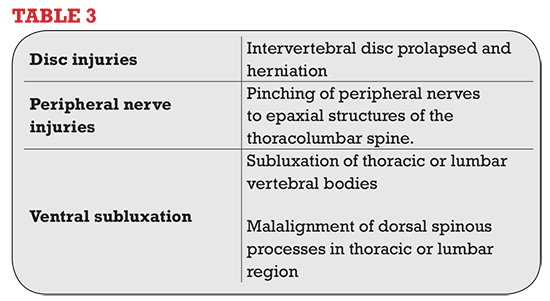
Table 3: Conditions that are alleged to cause back problems for which there is no scientific evidence
MALALIGNMENT OR DISPLACEMENT OF LUMBAR SPINOUS PROCESSES
This condition is often said to be a cause of back pain in horses, and is cited as a cause of chronic poor performance. It is claimed that horses have one or more spinous summits “put out” or laterally displaced, particularly in the caudal lumbar region. It is claimed that these vertebrae can be “put back” into their correct position by means of a manipulative technique that usually involves sharp pressure applied to the dorsal tip of the affect summit. From an anatomical point of view this claim is unacceptable as significant movement of the vertebrae or their spinous processes is impossible either naturally or with manual manipulation.
It is most likely that this malalignment of the back is an increase in postural tone of the affected muscles along the topline of the horse’s back (epaxial musculature). This is thought to be the cause of some orthopaedic deformities in people such as idiopathic scoliosis and club foot. Even though the situation in the horse’s spine is not strictly analogous to that seen in man, one can appreciate that differences in the loading of muscles on either side of the spine can occur in horses with chronic back pain. This may give some explanation for the sometimes dramatic and instant response to some chiropractic manipulations. These treatments presumably cause muscle relaxation and a subsequent improvement in performance. 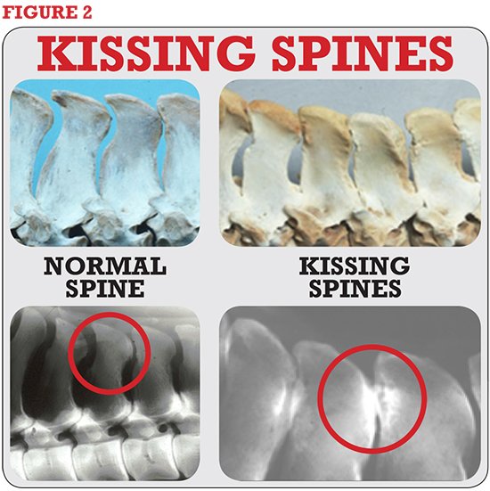
Figure 2: Radiographs and bony specimens from a normal horse and a horse with impingement of the dorsal spinous processes
OVERRIDING DORSAL SPINOUS PROCESSES (KISSING SPINES)
Inpingement and crowding of the thoracolumbar spinous summits has long been known as a cause of back pain in horses. However, the condition can also been seen in horses which do not show any signs of either poor performance or back pain. The result of this is that diagnosis of this condition is difficult, and requires careful clinical evaluation as well as taking high quality radiographs, performing a bonescan and an ultrasound examination. Horses affected with this condition are usually presented with a long history of poor performance, and they may have had many forms of empirical therapy. The condition occurs most commonly in young mature Thoroughbred or crossbred horses with short backs who are being used principally for eventing or showjumping. The impingement usually occurs underneath the saddle (T12-T17), but can occur in the caudal withers and cranial lumbar spine. The onset of clinical signs is usually insidious although a history of trauma from a fall is sometimes reported. These affected horses show increasing stiffness in the back, poor jumping ability, reduction in hindlimb impulsion and an unwillingness to work. A change in the horse’s temperament or resentment when being groomed or having the hind feet picked up by the farrier may also be seen. In severe cases the horse may be unwilling to roll or lie down. In chronic cases there may be minimal pain upon palpation, but the horse usually resents dipping the back and will have noticeable rigidity of the thoracolumbar spine on manipulation. On radiographs pressure points between opposing spines is seen as local bony reaction and small cyst-like areas of bone resorption, false joint formation is also often seen between the affected vertebrae.
Figure 3
FRACTURES OF THE THORACOLUMBAR SPINE
The most common fractures of this region are those that involve the dorsal processes of multiple vertebrae (broken withers). These are easy to diagnose as there is localised pain, heat and swelling in the region. The horse usually displays a stiff forelimb gait and stiff neck. The prognosis for these injuries is usually excellent, but healing of the fractures may take up to 6 months. Most horses are left with a depression over the withers, but there is no lasting effect on the horse’s performance.
Figure 3: Line drawing of a horse with fractured withers. Most of these injuries can be diagnosed via palpation as shown above.
Fractures of the vertebral bodies are usually associated with a fall or traumatic event and typically horses display paraplegia or profound neurological deficits. These fractures are invariably fatal.
We trust that this summary will help to illustrate the range of conditions that can affect the back in the performance horse, and also highlight some of the more common “myths” associated with this troublesome condition. The next article will focus on the treatment and management of back pain in the horse.
This article first appeared in the March 2013 issue of THM.